

Get new exclusive access to healthcare business reports & breaking news


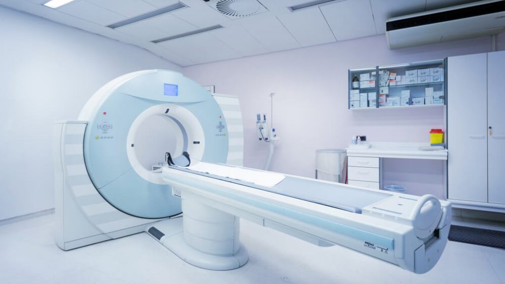
A CT (computerized tomography) or CAT (computerized axial tomography) scan creates a detailed image of the bones and organs within the body. It does this by combining data collected from a series of x-rays. The result of this data is a 2-dimensional image of a small portion of the area that the CT scanner viewed. The image view resembles a slice of the inside of the body and the data can also create a 3D image. Hospitals utilize this commonplace technology worldwide.
The machine that produces the CT scan is called a CT scanner. How it works is it sends several narrow beams through the body as it rotates around it in an arc. The completed CT scan is an intricately detailed picture of the region the beams entered. An x-ray machine, by comparison, emits a single radiation beam producing an image that is not nearly as detailed as is possible with a CT scan. For example, the x-ray detector of a CT scanner can see hundreds of density levels and can see tissues contained inside an organ.
The data produced by the CT scanner beams go to a computer that uses specific software to create a 3D cross-sectional image of the body region that the CT scan viewed. The final image appears on the computer screen for viewing. Depending on what the CT scan will examine, and the reason for the scan, there may be a need for a contrast dye. All this does is makes parts of the body in the scanner easier to see in the final image. CT scans are effective in aiding medical professionals with health diagnoses for their patients.
Because it can see things that an x-ray may miss, a CT scan is effective in many ways. It is the preferred method of diagnosing liver, lung, and pancreatic cancers. This is because the image details assist doctors with identifying the tumor’s location, size, and if it has spread to other tissue. A CT scan of the head reveals if there is bleeding, swelling, or a tumor present in the brain. It can uncover the presence of a tumor in the abdomen, or swelling of any kind in the liver, kidney, or spleen. Abnormal tissue, bone diseases, and damage to other skeletal structures are all easily detected with a CT scan.
It is common for some people to confuse a CT scan with an MRI (magnetic resonance imaging). Here is a look at both head-to-head. First, a CT scan employs x-rays. An MRI uses magnets and radio waves. An MRI can produce an image that shows ligaments and tendons, but a CT scan cannot. MRI technology is better for examining the spinal cord but CT technology excels at identifying cancer, brain bleeding, abnormalities in the chest, and pneumonia. An MRI produces better imaging for brain tumor detection. An organ injury is identified quicker with a CT scan as are broken bones and vertebrae. A CT scan also produces a sharper image of the lungs, and chest cavity organs. Because of the technology used, the CT scan machine price is high making it one of the most valuable tools in a modern hospital or clinic.
Depending on the scan, the patient may have to stop eating and drinking any liquid for a prescribed time before the scan appointment. Once at the appointment, the patient may have to strip down to their underwear and put on a supplied gown. The patient must remove all jewelry. Should there be a need for a contrast dye, it is provided either as an enema or injected intravenously. The patient will lie on a platform that will slide into a circular-shaped CT scanner machine. As x-ray images are taken, the platform the patient is lying on will move slightly to the next position. The patient must lie as still as possible through the entire scanning process which lasts only a few minutes.
A small, targeted dose of radiation is used in the average CT scan. The level is not harmful and has not been in individuals who have experienced multiple CT scans in their lifetime. The estimate of developing cancer from a CT scan measures at less than 1 in 2,000. As for the actual amount of radiation used, it equals about the same as what an individual would be exposed to over several years in the natural environment around them. It is important to note that CT scans are used only if a valid medical reason that requires one.
Although medical professionals have determined that the level of radiation used in a CT scan is not “significant enough to cause adverse effects in a developing embryo or fetus” they are not recommended for pregnant women. Any woman who suspects she may be pregnant should notify her doctor beforehand. The only time a pregnant woman should undergo a CT scan is if the benefits of the scan outweigh the risk. Breastfeeding mothers who require the contrast dye for a CT scan should avoid breastfeeding for 24 hours following the procedure. This is because the dye may end up in breast milk.
Because a patient must lie still on a platform that goes into the CT scanner machine, it may be stressful for those individuals who suffer from claustrophobia. In these cases, the patient should notify their doctor or the radiographer before the procedure. A tablet or injection will calm the patient before the scanning process can begin.
A CT scan is different from an x-ray and an MRI. Although all of these assist medical professionals in seeing inside the body, a CT scan is effective in many ways because of the added detail the multiple x-rays produce in the final image. There are times when an x-ray or an MRI is best suited, it just depends on what the doctor is trying to diagnose. If you require a CT scan, you can bet that your doctor has carefully weighed out the options and determined that a CT scan will assist them in getting to the root of your problem quickly and with more accuracy.